
Radial Annular Tear
Radial Annular Tear: General Information
A radial annular tear, often called a full-thickness annular tear, occurs when the annulus fibrosus develops a tear which extends from the nucleus, towards the periphery. As you may have already read on the discography page, there are several different classifications that are applied to annular tear.
 A grade I annular tear means that the tear extends from the nucleus into only the inner one third of the annulus fibrosus.
A grade II annular tear means the tear extends from the nucleus, all the way through the inner one third and into the middle one third of the annulus fibrosus.
A grade III annular tear means the tear extends from the nucleus, all the way through the inner one third, middle one third and into the outer one third of the disc, which we know is highly innervated by nociceptors (pain-carrying nerves), which is especially true in the posterior and posterolateral regions of the disc.
Therefore, in some people but not all, a grade III radial annular tear can become incredibly painful and even, in some but not all people, necessitate removal of the disc via lumbar interbody fusion.
Figure #2 demonstrates a full thickness, grade III, radial annular tear (usually just called a full thickness tear) of the disc. In this cartoon, a wicked inflammatory process has developed in the outer one third of the disc (red color) which was a pain generator in this patient.
Radial Annular Tears Versus Degeneration
Radial annular tears have been strongly associated with the development of degenerative disc disease. [3] So what does that mean? Well, as you have learned from the disc degeneration page, when the disc dries out it becomes more vulnerable to injury. Specifically, the lamellae are not as strong as they used to be and can more easily ripped through.
The degenerative/annular tear process begins with "cleft" formation within the innermost portion of the annulus fibrosus. As the disc dries out more and more lamellae rupture in the annular tear moves further and further away from the nucleus. And when it reaches the outer one third of the disc, and has the potential to generate significant low back pain. [3]
As described previously, sometimes radial annular tears will merge with concentric annular tear's in the periphery of the disc. The point where the two tears connect may be visible on MRI as a high-intensity zone (HIZ), which then makes the diagnosis of annular tear.
Radial annular tears: are they always painful?
It is important to understand that radial annular tears ARE NOT ALWAYS PAINFUL! This means that they can be found in people who are completely asymptomatic (do not have low back pain). [21]
Why some tears are painful and some aren't, is still not completely understood. The current theory states that if nuclear material migrates into the outer region of the annulus, which is well innervated with pain sensitive (nociceptive) nerve fiber, a chemical irritation and inflammation process occurs around these now exposed nociceptors (pain triggers at the beginning of sensory nerve fiber) and causes discogenic pain.
Another theory is that the degenerated and damaged nucleus pulposus can no longer support its share of the axial load of the body. Biomechanical studies have indicated that following an annular tear, axial load is no longer carried in the center of the disc but instead shifts posteriorly right over the nerve-infested posterior and posterolateral annulus fibrosus. Therefore, in addition to a painful inflammatory reaction, now you get a mechanical (a grinding) irritation of those already inflamed and irritated nociceptive fibers. Another theory is that an autoimmune reaction occurs when the virgin nucleus pulposus cells get into the posterior annulus.
Annular Tears: Can They Heal?
Nobody really knows for sure how much an annular tear can heal. They are impossible to study in vivo and we must relying cadaver studies to answer this question. With that caveat in mind, it appears that scar tissue can form, at least in the outer one third of the disc, and seal that portion of the tear shut. But what about the rest of the tear? Cadaver studies indicate that angiogenesis (the formation of new blood vessels) does occur and follow the annular tear all the way down to the center of the disc. This is a double-edged sword, however! Although the new blood vessel formation will allow scar tissue to form all the way down the tear and possibly seal it, where blood vessels go, so do nerves. Therefore, it's not surprising that cadaver studies have demonstrated nerve fiber with nociceptive capability bleeding all the way into the center of the disc in patients with full thickness annular tear. So what does that mean? It means that patients who have suffered a full-thickness annular tear are now more "wired" for pain and may be able to experience pain when normal people don't. In other words they are more delicate and more prone to flare up. This would explain why one somebody seriously injures a disc, there usually "in the club" so to speak, for they are apt to have at least a few flareups per year for the rest of their lives.
What about healing time? Again, we have no research on this, but anecdotally speaking, symptomatic annular tear can take many months and even many years to heal to the point where the patient becomes relatively asymptomatic.
There is another anecdotal rule of thumb that I should put forth: patients who demonstrated an annular tear and MRI usually get better within four months. However, if they are not better within four months, they are typically still suffering at 18 months! The good news is that typically these patients are doing better at the 4-5 year mark. Unfortunately, some people just can't wait that long – especially people with arduous work.
radial annular tears: classification of
As talked about previously, radial annular tears have several different classes of evolution. These classes were thoroughly described on the discography page. So if you're interested, please visit that page for more information.
Internal Disc Disruption: (aka: IDD)
 When full thickness annular tears are thought to be symptomatic and the cause of the patient's pain syndrome, a diagnosis of internal disc disruption (a.k.a. internal disc derangement, or IDD) is sometimes put forth. Therefore, IDD is nothing more than a symptomatic full thickness, or any other type of annular tear.
Instead of using the term IDD, many practitioners, however, use the term "discogenic pain syndrome" for the diagnosis. They both me the same thing.
Again, the only way to really visualize an annular tear is through the discography procedure.
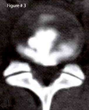
The image left (figure #3) demonstrates a perfect example of a grade IV annular tear, as visualized on CT after the performance of discography. Note the ship-anchor appearance on the axial image.
Because the patient suffered severe low back pain and some radiating lower extremity pain during the pressurization part of discography, (in other words, he suffered concordant pain) he was given a diagnosis of internal disc disruption and lumbar interbody fusion surgery was scheduled. Why interbody fusion? It is believed that you must remove the inner portion of the disc and then take all pressure off the disc by placing an interbody spacer (cage) in place of the disc. Please go to my fusion page to learn more about this.
History and Research:
IDD was first described by Crock in 1970 (8) and again in 1986 (9). They described it as a ‘disruption of the internal architecture of the disc without signs of disc protrusions or without positive signs for nerve root compression’.
In 2003, Lee et al. reviewed the research on IDD from 1985 through 2000 (10). They summarized that of the 13 research papers on the subject, there was not much agreement on the subject. There was some agreement on what constituted a diagnosis of IDD. Here’s the factors that most researcher’s felt constituted a diagnosis of IDD: Lower back pain; a painful disc on provocative discography (concordant pain is even better); and a normal neurological examination (i.e. no loss of reflexes, no loss of muscle strength or atrophy, and no sensory loss). Other criteria for the diagnosis of IDD were not so universally agreed upon, including the presents of an HIZ sign on MRI, degeneration, desiccation, and a history of trauma. Based on their review of 15 years worth of research, Lee et al. boldly concluded that “IDD is not real, but a hypothetical disease”. This Korean group further stated “Our personal view is that IDD is a doctor-made disease, that is, [sic] an Iatrogenic Disc Disorder, which may lead to an unconventional invasive operation.”
Therefore, it is not surprising that the diagnosis of IDD was and continues to be controversial. We need more research on the subject before more universal acceptance of the diagnosis can be reached.
Rim Lesions | Concentric Tears | HIZ Sign | Anular Tear Home
References:
2) Schmorl G, Junghans H, “The human spine in health & disease”. New York: Grune & Stratton, 1971
3) Osti OL, Vernon-Roberts B, et al. “Annular Tears & Disc Degeneration” J Bone Joint Surg [Br] 1992; 74-B:678-82
5) Osti OL, et al “Volvo Award- Anulus Tears & Intervertebral Disc Degeneration – Spine 1990; 15(8):762-766
7) Aprill C, Bogduk N, “High Intensity Zone” - The Brit Jour Radio 1992; 65, 361-369
8) Crock HV, “A reappraisal of intervertebral disc lesions” – Med jour Australia 1970; 1:983-989
9) Crock HV, “Internal Disc Disruption” – Spine 1986; 11:650-653
10) Lee KS et al. “Diagnostic criteria for the clinical syndrome of IDD” - Brit Jour Neurosurgery 2003; 17(1) 19-23
14) Sachs BL, et al. “Dallas Discogram Description a New Classification of CT/Discography” – Spine 1987; 12(3):287-294
15) Schellhas KP, et al. “Lumbar Disc High-intensity Zone – MRI & Discography” – Spine 1996; 21:79-86
21) Boos N, et al. “Volvo Award in Clinical Science: The Diagnostic Accuracy of MRI” – Spine 1995; 20(24):2613-2625
33) Moore RJ, “The Origin & Fate of Herniated Lumbar IVD Tissue.” Spine – 1996;21:2149-2155
© Copyright 2002 – 2015 by Dr. Douglas M. Gillard DC - All rights reserved